Published March 16, 2018
Until the 19th century, the relationship between the function and the physiology of the nervous system was largely a mystery. Physicians believed in the vital importance of the brain but knew little about its structure and purpose. For hundreds of years, conventional wisdom in medicine followed the second-century physician Galen in holding that certain pneumata, or bodily spirits, were stored in hollowed-out cavities in the brain (the anterior ventricles), and those spirits’ motion around the body was necessary for seeing, hearing, tasting, and feeling.
Neuroscientific understanding made rapid strides in the 1800s. The French-men Paul Broca, a surgeon, and Jean-Baptiste Bouillaud, a physician, both succeeded in linking specific parts of the brain to specific functions—for example, there is a part of the brain (now called Broca’s area) that is known to be associated with speech.
But what was happening at the microscopic level? Sure, some locations in the brain seemed to control language, but what did that mean? Do cells send signals to initiate speech? What do those cells look like and how might they be connected? Answers to these questions would provide a huge leap forward in the progress of neuroscience.
That leap is in part attributable to the Spanish physician-artist Santiago Ramón y Cajal. Born in 1852, Cajal was the troublemaking son of a professor of anatomy; the father eventually persuaded the artistically inclined son to study medicine. Cajal served as a medical officer in the Spanish Army and eventually became an anatomy professor himself, conducting research focused on the nervous system. In order to elucidate the workings of the brain, he improved upon an existing cell-staining technique and then studied neurons (brain and nerve cells) under the microscope. Cajal found that spaces exist between neurons—what we today call synapses—and recognized certain patterns in the way neurons line up next to each other. Consequently, he could tell in which direction along the cell signals traveled.
The importance of these two discoveries cannot be overstated. Treatments available for seizures, autoimmune diseases, depression, schizophrenia, anxiety, and more depend on the synapses between neurons; drugs act on receptors and chemicals in this space. Moreover, diagnostic tests for nerve damage lean on our knowledge of the direction of neuronal signals. It is for these discoveries that Cajal was awarded the Nobel Prize in Physiology or Medicine in 1906. And his ability to sketch what he saw when he peered through his microscope aided his research immensely.
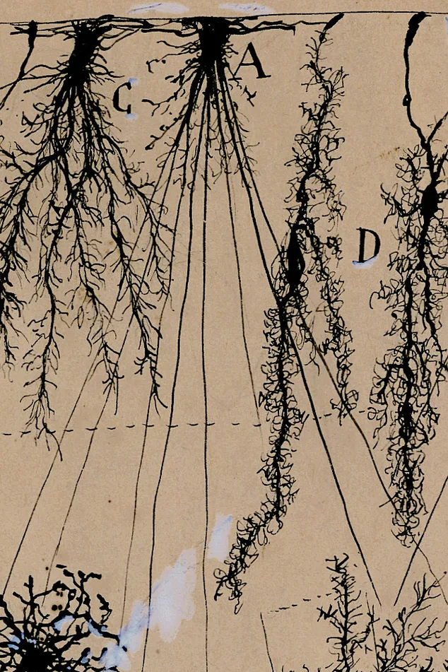
Now, 80 of Cajal’s drawings are on display in an exhibition at New York University’s Grey Art Gallery. They are mesmerizing. One of Cajal’s most famous illustrations shows a Purkinje neuron from the human cerebellum (the part of the brain involved in coordination); dozens of root-like projections emerge from the cell body, filling the entire page, searching for their companion cells beyond the borders of the sheet as they sway on the parchment. Cajal wrote of this neuron, “In our parks are there any trees more elegant and luxurious than the Purkinje cell from the cerebellum . . . ?”
In another image, a white blood cell migrates across a blood vessel in successive stages, the corpulent cell projecting tentacles to the vessel wall, burrowing through it and pulling itself over to the other side, misshapen but otherwise unharmed. Of an event like this, Cajal wrote in his autobiography, “I remember that once I spent twenty hours continuously at the microscope watching the movements of a sluggish leukocyte in its laborious efforts to escape from a blood capillary.”
There is more than just beauty in these illustrations. Cajal meant for them to appear with his scientific publications. He labeled them with letters or arrows to point out structures of interest. They remain so accurate that many will appear familiar to anyone who has perused recent neuroanatomy textbooks. Moreover, some parts of the images seem to have been playfully inserted by their creator. In one sketch of cells from the cerebral cortex of a man suffering from paralysis, a ghoulish face replaces the body of a neuron and peers down at the other damaged cells.
Despite the wonderful drawings on display, it is hard to make sense of each image’s purpose in the exhibition’s larger story. Although it is divided into sections—“Cells of the Brain,” “Sensory Systems,” “Development and Pathology,” “Neuronal Pathways,” and “Seeing the Beautiful Brain Today”—these divisions seem somewhat arbitrary. Why are these considered separate sections? After all, sensory systems extend into brain cells, and development and pathology occur in brain cells. And what’s on offer in the sections doesn’t differ all that much. In the first section, one can view a drawing of a pigeon’s Purkinje neuron; in the second, one can see the retina of the lizard. The exhibition neglects to guide the visitor with a coherent narrative.
Furthermore, the last section, “Seeing the Beautiful Brain Today,” feels stilted and out of place. It contains more specialized images made possible by today’s tools and techniques—magnetic resonance imaging (MRI), electron micrography, and so on—which offer remarkable ways of viewing the brain as a whole or in various parts. These are invaluable for modern medicine and some of them are quite striking. But as images, as objects worth beholding and critiquing for their aesthetic value, they pale in beauty relative to Cajal’s pencil-and-paper works. * * *
In his book Advice for a Young Investigator, Cajal argues that art and science should be happily merged: The investigator ought to possess
an artistic temperament that impels him to search for and admire the number, beauty, and harmony of things; and—in the struggle for life that ideas create in our minds—a sound critical judgment that is able to reject the rash impulses of daydreams in favor of those thoughts most faithfully embracing objective reality.
Artistic and scientific ways of thinking naturally balance each other, Cajal claims. But art without science is dissatisfying, subjective, and ephemeral:
Art depends on popular judgments about the universe, and is nourished by the limited expanse of sentiment. . . . In contrast, science was barely touched upon by the ancients, and is as free from the inconsistencies of fashion as it is from the fickle standards of taste. . . . And let me stress that this conquest of ideas is not subject to fluctuations of opinion, to the silence of envy, or to the caprices of fashion that today repudiate and detest what yesterday was praised as sublime.
This is a chilling and vicious attack on art practiced without the sciences. And it is shortsighted to make such an accusation about the arts when the scientific enterprise is, despite Cajal’s optimism about it, sometimes subject to the same caprices.
Still, if he romanticized science at the expense of art, Cajal was certainly right about the potential to merge the two. His drawings exemplify this. They go hand-in-hand with his discoveries, beautifying and clarifying them.
Yet they didn’t clarify everything. For all our knowledge about the nervous system and for all our high-tech imaging capabilities, even today we must sometimes feel like Cajal or even Galen: ignorant, overwhelmed, and saddened by what we still don’t know about, for example, incurable nervous-system afflictions like Lou Gehrig’s disease or Parkinson’s disease. We must hope that future creative insights—perhaps via a marriage of art and science like that achieved by Cajal—will push back against these terrible unknowns.